Crusted sores on nose


An 18-year-old girl asked her family physician to take a look at the crusted sores on her nose and the rash on her face—both of which had developed a week earlier. She was so upset by the sores on her nose that she was covering her face with a kerchief. She had been previously healthy and had no fever, chills, or night sweats. She had no history of trauma or sunburns. Her only symptom was some mild tenderness of the affected skin.
What's your diagnosis?
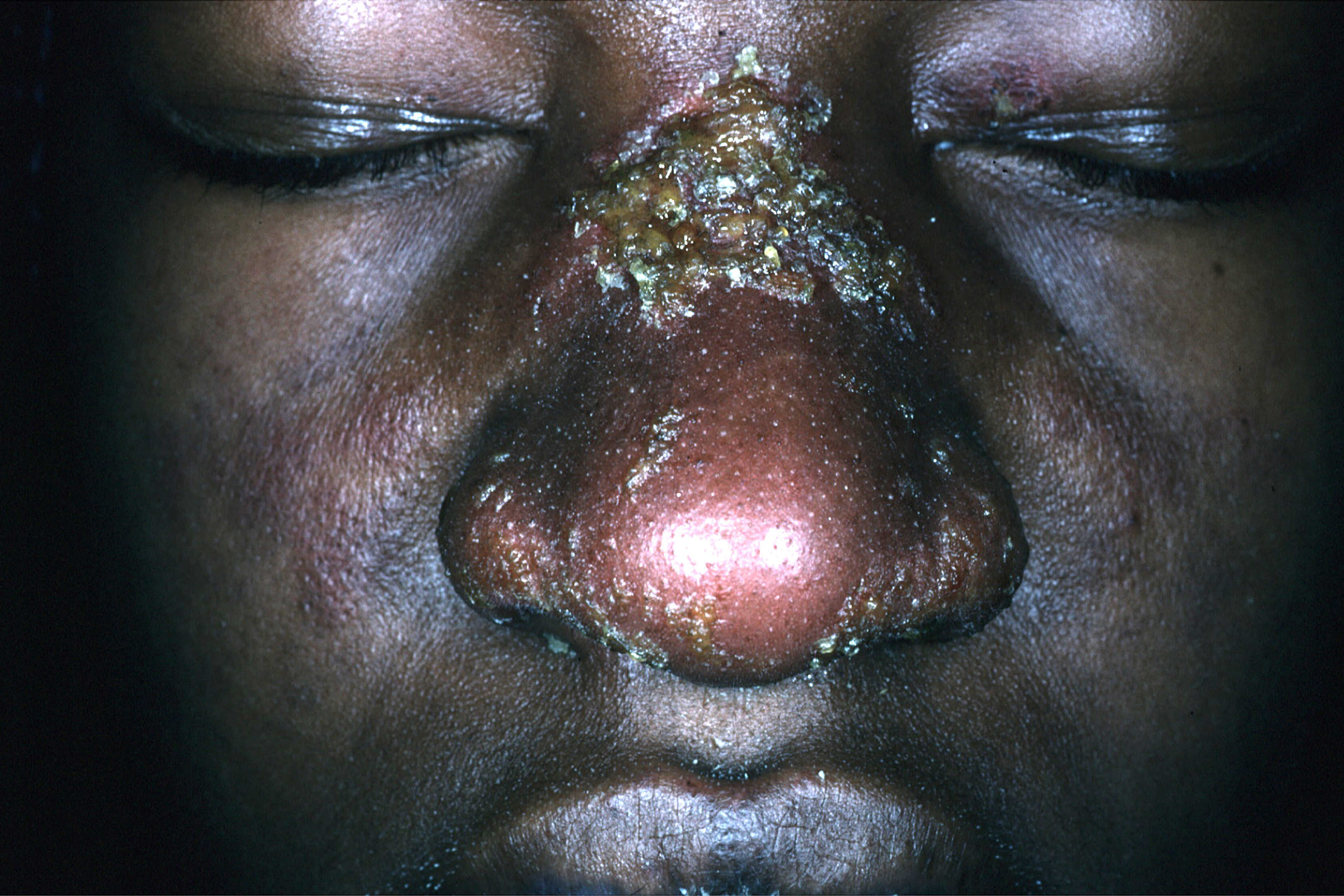
The family physician initially diagnosed impetigo (based on the honey crusts), but it soon became clear that she was also suffering from systemic lupus erythematosus (SLE).
The physician initially treated the patient with oral cephalexin 500 mg twice daily for 10 days and the crusting resolved. However, since there was erythema over the malar area, an antinuclear antibody (ANA) test was ordered. The ANA test was positive at a 1:160 dilution. A homogeneous nuclear pattern was present, as is commonly seen in SLE and drug-induced lupus. The comprehensive chemistry panel and complete blood count were normal. At the time, she did not meet criteria for SLE. However, 6 months later she developed severe lupus cerebritis requiring a prolonged hospitalization.
The diagnosis of SLE is made if 4 or more of the manifestations mentioned below are present (either serially or simultaneously) in the patient at the time of presentation or were present in the past.
- Systemic symptoms such as low-grade fever, fatigue, malaise, anorexia, nausea, or weight loss.
- Arthralgias that are usually out of proportion to physical findings. Polyarthritis is symmetric, nonerosive, and usually nondeforming. In longstanding disease, rheumatoid-like deformities with swan-neck fingers are commonly seen.
- A malar or butterfly rash over the cheeks and bridge of the nose, sparing the nasolabial folds.
- Rash associated with photosensitivity to ultraviolet light.
- A discoid rash consisting of erythematosus raised patches with adherent keratotic scaling and follicular plugging. Atrophic scarring may occur in older lesions.
- Ulcers (usually painless) in the nose, mouth, or vagina.
- Pleuritis (as evidenced by a convincing history of pleuritic pain or rub) or evidence of pleural effusion.
- Pericarditis as documented by EKG, rub, or evidence of pericardial effusion.
- Renal disorder such as cellular casts or persistent proteinuria >0.5 g/d or >3+ if quantitation is not performed.
- Central nervous system symptoms ranging from mild cognitive dysfunction to psychosis or seizures.
- Hematologic disorders such as hemolytic anemia, leukopenia (<4000/mm3 total on ≥2 occasions), lymphopenia (<1500/mm3 on ≥2 occasions), or thrombocytopenia (<100,000/mm3 in the absence of precipitating drugs).
- Gastrointestinal symptoms that may include abdominal pain, diarrhea, and vomiting.
- Vasculitis, which may be severe and can include retinal vasculitis.
- Immunologic disorders, such as a positive antiphospholipid antibody, anti-DNA, anti-Sm, or a false positive serologic test for syphilis (known to be positive for at least 6 months and confirmed by a negative treponema specific test).
Photos and text for Photo Rounds Friday courtesy of Richard P. Usatine, MD. This case was adapted from: Mayeaux, EJ. Lupus erythematosus (systemic and cutaneous). In: Usatine R, Smith M, Mayeaux EJ, et al, eds. The Color Atlas of Family Medicine. New York, NY: McGraw-Hill; 2009:766-771.
To learn more about The Color Atlas of Family Medicine, see:
* https://www.amazon.com/Color-Atlas-Family-Medicine/dp/0071474641
* https://www.mhprofessional.com/product.php?isbn=0071474641