
A Rare Finding of Simultaneous Lung Cancers


Background: Lung cancer is the leading cause of cancer death in the United States. Although rare, some cases present with multiple primary lung cancers (MPCs), defined as the occurrence of > 1 primary cancer simultaneously or sequentially in the lung. This report highlights the challenges of diagnosis and treatment associated with MPC.
Case Presentation: A male aged 72 years presented to the Veterans Affairs Caribbean Healthcare System emergency department with shortness of breath and dyspnea on exertion. Chest computed tomography revealed a new large left pleural effusion. The pleural effusion analysis with pathology was remarkable for lung adenocarcinoma. A pleural biopsy pathology found simultaneous spindle cell carcinoma and lung adenocarcinoma.
Conclusions: This case is a rare instance of concomitant lung adenocarcinoma and spindle cell carcinoma.
Lung cancer is the second-most common cancer and the leading cause of cancer death in the United States.1 A significant number of patients with lung cancer have advanced disease at clinical presentation. Some patients present with multiple primary lung cancers (MPCs), defined as the occurrence of > 1 primary cancer simultaneously or sequentially in the lung.2 Simultaneous primary lung cancer is extremely rare, occurring in about 0.01% to 0.54% of individuals screened for lung cancer.3 Spindle cell carcinoma of the lung is a rare and aggressive type of non-small cell lung cancer characterized by spindle-shaped tumor cells, which contributes to challenges in diagnosis and treatment.
Case Presentation
A 72-year-old male former smoker with a medical history of prostate adenocarcinoma presented to the Veterans Affairs Caribbean Healthcare System emergency department with progressive dyspnea on exertion and cough. His vital signs were remarkable for hypoxemia with an oxygen saturation of 88% on room air. A chest computed tomography (CT) revealed a large left-sided pleural effusion with associated abnormal pleural nodularity concerning for malignant effusion. A left-sided 10.2 Fr chest tube was successfully inserted and pleural fluid sample were collected. The pleural effusion analysis revealed pH 7.37, 1169 pleural cell count, malignant cells, 5.0 pleural protein, and 301 U/L lactate dehydrogenase (LDH). Serum protein was 6.5 and LDH 190 U/L. Light criteria were consistent with an exudative effusion. The pleural pathology was consistent with malignant pleural effusion, with positive immunostains including pankeratin, p53, and calretinin.
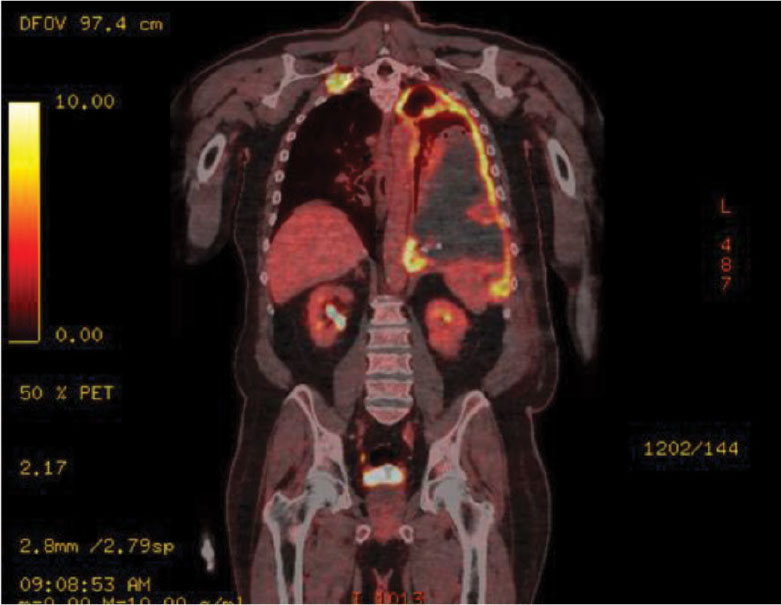
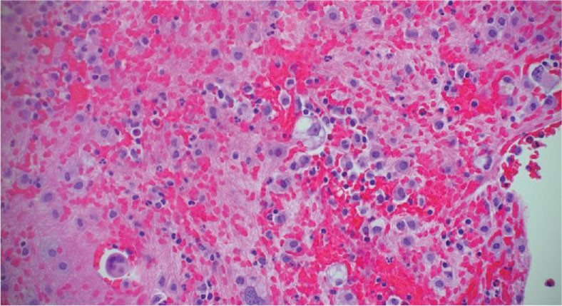
The patient’s pathology report was initially concerning for mesothelioma; however, he reported no risk factors for asbestos exposure. For further evaluation and staging of malignant mesothelioma, a positron emission tomography/CT was conducted, which was remarkable for multiple hypermetabolic lesions, including left and right thoracic pleura, and bilateral mediastinal lymph nodes (Figure 1). Hematology and oncology services requested pleural based biopsy. Pleural biopsy revealed a malignant neoplasm consisting of spindle cells, epithelioid cells, and rare giant cells with positive immunostains including pankeratin, TTF1, AE1/AE3, and thyroid transcription factor 1 (TTF-1) (Figure 2). These findings were diagnostic for pleomorphic carcinoma, which in this sample included poorly differentiated high-grade pulmonary adenocarcinoma and spindle cell carcinoma.
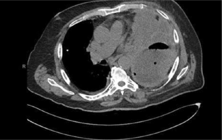
The patient’s hospitalization was complicated by superimposed pneumonia. Another chest CT revealed new findings of air fluid collection and loculated effusion (Figure 3). The patient’s laboratory results were remarkable for increasing leukocytosis and procalcitonin. New pleural fluid analysis revealed a pH of 7.21, cell count pleural of 1169, pleural protein of 2.6, LDH > 2500, and glucose < 2 mmol/l. The patient’s serum protein was 4.4 and his LDH was 208 (Figure 5). Light criteria were consistent with exudative effusion again. This finding was highly suggestive of complicated parapneumonic pleural effusion for which medical therapy with broad spectrum antibiotic therapy and alteplase and deoxyribonuclease were administered.
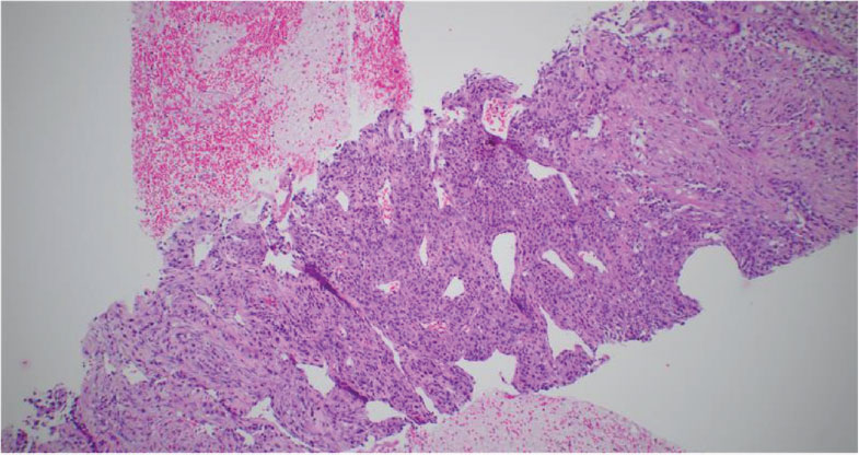
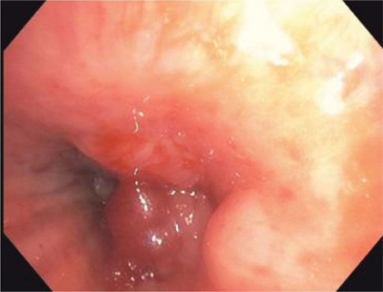
Despite therapy, the patient continued to deteriorate and developed respiratory distress and hypoxemia requiring endotracheal intubation and mechanical ventilation. While in the intensive care unit, the patient underwent fiberoptic flexible bronchoscopy for further evaluation, which revealed left mainstem endobronchial lesion and pathology positive for a poorly differentiated malignant neoplasm (Figure 6). Positive immunostains had presence of CK7, pankeratin, and WT1. The patient was subsequently determined to have concomitant lung adenocarcinoma and spindle cell carcinoma. Given the poor prognosis, family members opted for palliative care.
Discussion
The rate of misdiagnosis of MPC through biopsy specimens is high. Obtaining an adequate tissue sample and performing a thorough pathologic evaluation are essential for accurate diagnosis. The clinical impact of combined non-small cell lung carcinoma with spindle cell tumor remains unclear. Non-small cell lung cancer comprises about 80% to 85% of all new lung cancer diagnoses.4 Lung adenocarcinoma is the most common lung cancer in the US and categorized as non-small cell lung cancer.5 Typically, lung adenocarcinoma demonstrated positive immunostaining for TTF-1, napsin A, and keratin 7.6
The incidence rate of spindle cell carcinoma is about 0.4% of all lung malignancies, demonstrating its rarity.2 Diagnosis can be delayed given low incidence of malignancy and complexity in accurate diagnosis. Patients diagnosed with spindle cell carcinoma have poor prognosis, and overall survival time is poor. Pleomorphic carcinomas have a high incidence of PD-L1 expression (60% to 90%). Immune checkpoint blockades (ICBs) can significantly improve the prognosis of spindle cell carcinoma compared with chemotherapy. However, most patients who receive ICB with a clinical response experience relapse because of acquired resistance. The prognosis of pleomorphic carcinoma is worse than that of other non-small cell lung cancers. Therefore, additional therapeutic strategies are needed to improve outcomes for this aggressive malignancy.
Conclusions
Simultaneous primary lung cancer is extremely rare and occurs in < 1% of individuals screened for lung cancer. The rate of misdiagnosis of MPC through biopsy specimens is high. This case highlights the challenges of diagnosis and treatment associated with multiple simultaneous primary lung cancers.
